Eyelid Skin
The skin is thin but, typical of all skin, has fine hairs and sweat glands. The loose subcutaneous tissue, with collagen and elastin fibers, contains small quantities of fat and some granules of yellow or brown-colored material.
The layers of structure of the eyelid, from front to back are:
- Skin
- Subcutaneous tissue
- Orbicularis oculi muscle
- Layer of fatty tissue
- Tarsal Plate and tendon of Levator palpabrae superioris
- Meibomian glands
- Conjunctiva
- Eyelashes and glands of Moll at the margins
The orbicularis oculi muscle is formed from a flat sheet of circular fibers. It is described by anatomists in three portions, though they function together: orbital, palpebral and lacrimal. The orbital portion is attached at the medial orbit to frontal and maxillary processes and to the medial palpebral ligament of the tarsal plate; the fibers encircle the orbit, blending superiorly with occipitofrontalis and corrugator. The thin palpebral portion is attached to the medial aspect of the orbit, to the medial palpebral ligament, and passes to form a circle of fibers anterior to the orbital fascia, anterior to the tarsal plate within the eyelids, and merges with the lateral margin of the eye; upper and lower fibers interlace in a commissure as the lateral palpebral raphe. A distinct portion of ciliary bundle of Riolan muscle fibers are located behind the eyelashes.
A layer of fatty tissue, the palpebral fascia of variable thickness, lies between the muscle and the tarsal plate and levator tendon attached to it. In this lie blood vessels and some strands of smooth muscle, the superior tarsal muscle of Müller.
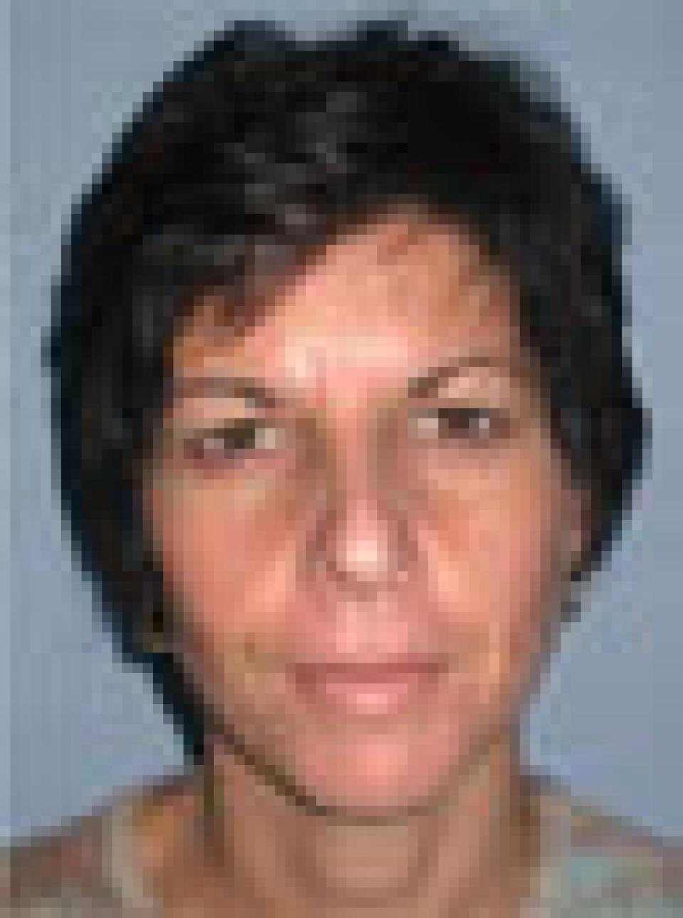 before
 after
after
This case depicts a 45 year old woman who wished to address her "heavy" upper eyelids and the fact that others often perceived her as angry.
Dr. Belsley felt that she would benefit from elevating the position of her eyebrows in addition to having upper and lower eyelid lifts. The combination of the brow lift and eyelid lifts opened up her eyes and defined her.
Upper eyelid creases, while maintain a youthful fullness to the upper eyelid. The post-operative photographs depict her appearance at two weeks after surgery. Dr. Belsley performs brow lifts using a few short incisions within the hairline. The position of the eyebrows alone can be elevated using the incision through which an upper eyelid lift is performed.
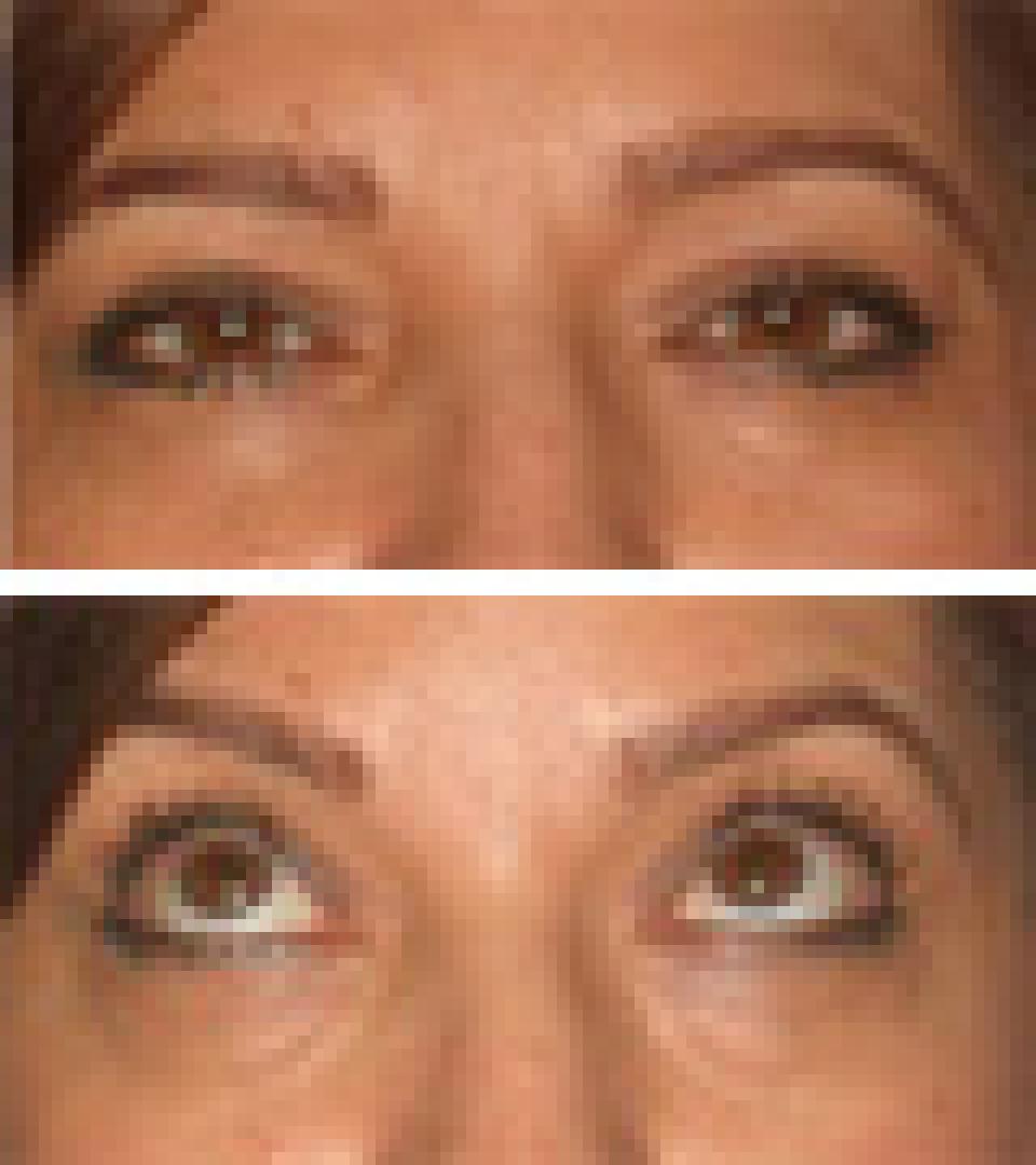 before
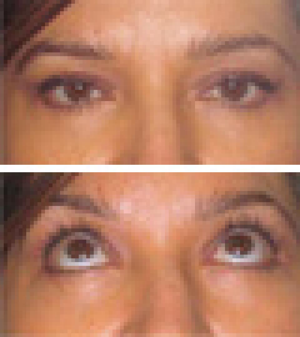 after
after
This case depicts a 44 year old woman who underwent a lower eyelid lift in which the lower eyelid fat was rearranged rather than removed.
The result of this procedure is a smooth lower eyelid contour devoid of the bulging or "bags" that many individuals exhibit. The change can most readily be seen in the photographs in which an individual is looking upwards.
The post-operative photographs depict this woman's appearance approximately four months after surgery.
 before
 after
after
This case depicts a 56 year old woman who wished to improve the appearance of sagging skin that she had developed as a result of age and dramatic weight loss.
She underwent an endoscopic brow lift, upper and lower eyelid lifts, a short scar face lift and a neck lift. Her post-operative photographs depict her appearance at approximately 4 months after surgery.
Although a properly executed short scar face lift can effect some improvement in the appearance of sagging skin in the neck with no incisions behind the ears, individuals with an extensive amount of skin and muscle descent require longer incisions and a formal neck lift to achieve dramatic results.
 before
 after
after
This case depicts a 55 year old woman who wished to achieve a more youthful facial appearance. In addition, others felt that she often appeared "angry." She underwent an endoscopic brow lift, upper and lower eyelid lifts and a short scar face lift which addressed the skin and fat beneath the chin, the jowls and elevated the midface. The post-operative photographs depict her appearance approximately three and a half months after surgery.
A short scar face lift often requires no incisions behind the ears and can achieve an excellent result in appropriately selected cases. The recovery time ranges from two to four weeks, although some individuals return to work as early as one week after surgery.