Midface
The total zone between eyes and mouth is known to the anatomists by this term, but the surgeon is more probably thinking of the cheeks and not the nose.
It is a region of great surgical consequence related to congenital malformations, as well as the results of trauma and aging.
The underlying skeletal structure is dominated by left and right maxilla bones, fused in the center and forming not only the upper jaw but also the bulk of the cheek bones. The maxilla extends upwards to participate in forming the nasal wall and floor of the orbit, and extends its process laterally to join with the zygoma bone to form the arch of the cheek.
Slightly below the inferior orbital margin, the infraorbital foramen is traversed by the trigeminal nerve which supplies sensation to the face and teeth (the facial nerve supplies motor power).
At the point where the two maxillae join in the midline beneath the nasal cavity they form the nasal spine, at the upper lateral aspect of the bridge of the nose they articulate with the small nasal bones.
There is a continuous layer of muscles in the face, the “superficial muscle aponeurotic system,” more conveniently shortened to SMAS, but certain groupings of muscle fibers are recognized, function as a unit with specific actions and are appropriately named.
The orbicularis oculi muscle forms a ring of muscle fibers around the eye; of its three parts, it is the lower fibers of the outer section, named orbital portion, which lie over the maxilla, inferior to the orbit, and are attached in part to the frontal process of the maxilla. The muscles that dilate or compress the nostrils are laterally attached to the maxilla.
The triangular, fibrous and fatty, Malar Fat Pad lies along the edge of the nasolabial fold. Its upper border covers the orbital rim and the orbital part of the orbicularis oculi. It lies beneath the skin and subcutaneous fat, superficial to the SMAS but not attached to it. As a person ages, the malar fat pad slides inferior and medial to its original position, and deepens the nasolabial fold.
 before
 after
after
This case depicts a 52 year old woman who underwent an upper eyelid lift and a short scar face lift.
Her post-operative photographs depict her appearance at approximately four months after surgery.
Although a short scar face lift does not require any incisions beneath the ears, it can have a profound effect on the jowls and the skin and fat beneath the chin.
 before
 after
after
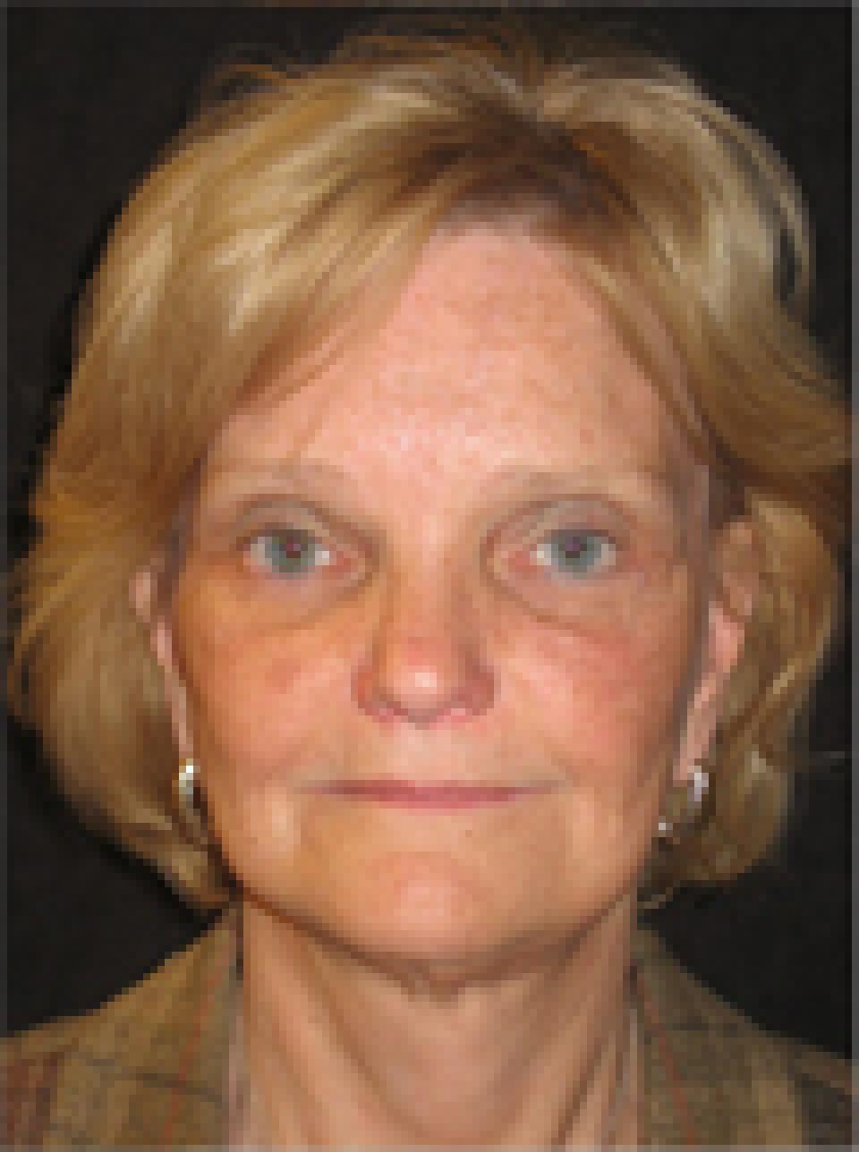
This case depicts a 56 year old woman who wished to improve the appearance of sagging skin that she had developed as a result of age and dramatic weight loss.
She underwent an endoscopic brow lift, upper and lower eyelid lifts, a short scar face lift and a neck lift. Her post-operative photographs depict her appearance at approximately 4 months after surgery.
Although a properly executed short scar face lift can effect some improvement in the appearance of sagging skin in the neck with no incisions behind the ears, individuals with an extensive amount of skin and muscle descent require longer incisions and a formal neck lift to achieve dramatic results.
 before
 after
after
This case depicts a 58 year old woman who wished to achieve a more youthful facial appearance.She underwent upper and lower eyelid lifts and a short scar face lift which addressed the skin and fat beneath the chin, the jowls and elevated the midface.
Her post-operative photographs depict her appearance at approximately nine months after surgery.
I try to perform a "mid-face lift" or "cheek lift" as a routine part of a short scar facial rejuvenation procedure. In individuals who have full cheeks that have descended over time, this portion of the procedure complements an aggressive correction of the jowls.