Ventral Hernia
The Latin for belly is venter (also meaning the womb), and a hernia occurs when some substance that should be completely contained manages to escape through its containing wall. A ventral hernia is therefore one that occurs in the belly, and in practice does not include umbilical nor inguinal herniae but is used for the midline of the abdomen.
The two rectus abdominis muscles are contained within a sheath of dense fascia, confluent with the aponeurosis (flat tendon) of the internal and external oblique abdominal muscles. Between the muscles, at what is called the linea alba, the sheath is less dense. It is easy to palpate the gaps between the muscles, particularly at the upper end, close to the chest.
When opening the abdomen (laparotomy) most surgeons will make their incision to the side of the midline, cutting through anterior and posterior walls of the rectus sheath, so they can be separately sutured and less likely to come apart than an (easier) incision made through the linea alba.
When the abdomen expands, as in pregnancy and obesity, the rectus muscles tend to get stretched apart, widening the gap between them (divarication). After delivery, abdominal strengthening exercises will restore normal muscle tone and position to the rectus muscles. But as obesity progresses, as the rectus muscles are stretched further apart, the linea alba becomes thinned. In such a patient, as he lies on his back and raises his head and shoulders, increasing intra-abdominal pressure, a ridge of ventral hernia protrudes between the rectus muscles. In itself this is not dangerous. Such a hernia is extensive, does not get trapped and there is no fear of strangulation or bowel obstruction that may be a concern in umbilical and inguinal herniae.
Although more properly called an incisional hernia the term of “ventral hernia” may also be applied to the type where an old incision has weakened and the abdominal contents protrude through the gap in the abdominal wall. Commonly this occurs when the patient has both gained substantial weight and lost abdominal muscle strength. This type of hernia may have a tighter orifice, a harder mouth and is potentially dangerous.
Repairs of incisional herniae are usually considered necessary, and may be performed by open or laparoscopic surgery, with or without the use of a reinforcing mesh.
Repairs of ventral herniae from divarication of the rectus muscles are usually considered optional, are not likely to be undertaken unless the patient has lost weight, and more commonly are performed in conjunction with an abdominoplasty after bariatric surgery.
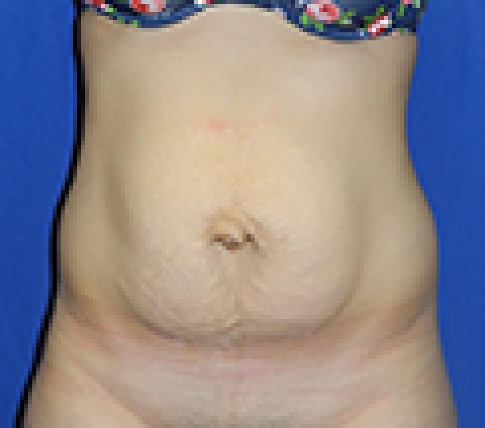
 before
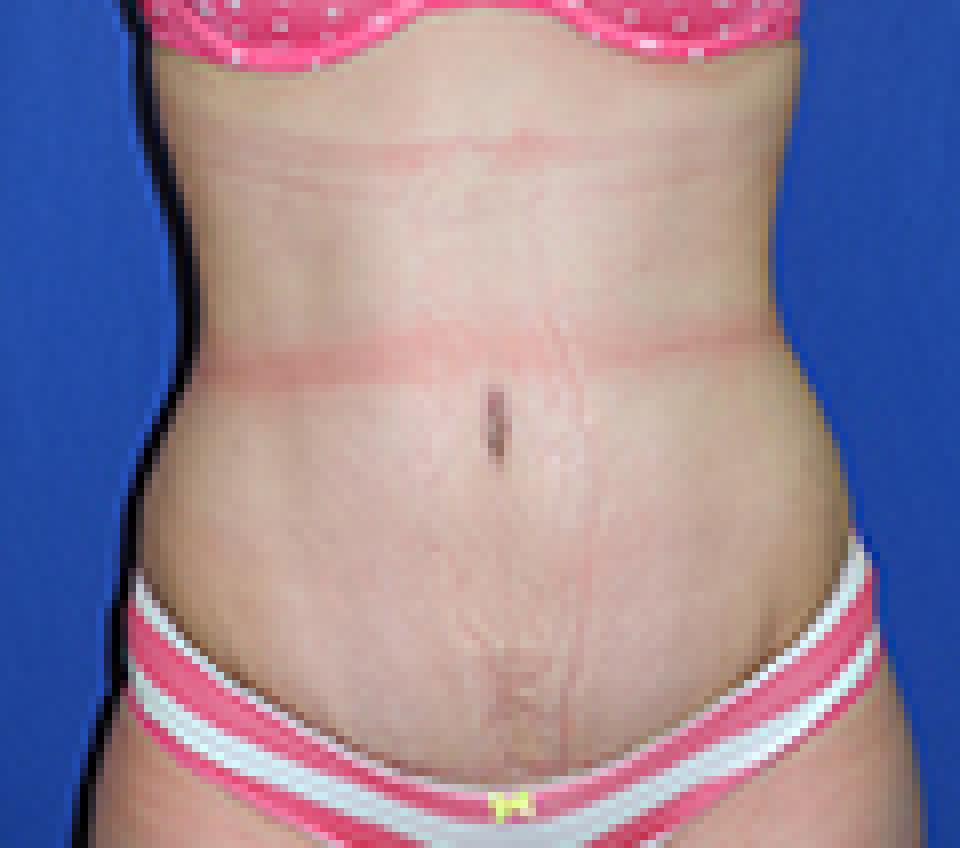
 after
after
This is a 21 year old woman with a BMI of 25.5 who had borne one child and became very displeased with the contour of her abdomen as a result. She had already been told that she had an umbilical hernia and wished to undergo repair of both issues at the same time.
She underwent lipoabdominoplasty, a procedure that treats the front of the abdomen by safely combining liposuction and abdominoplasty, along with liposuction of the bilateral anterior flanks, which refers to the front half of the "sides" of the body. Her umbilical hernia was repaired at the same time by Scott Belsley, MD, FACS, a board certified general surgeon, using a biological mesh that is incorporated into the body's own tissues over time. Dr. Scott Belsley and I often collaborate on cases that involve both plastic surgical and general surgical issues.